
Where is an ant’s heart?
An ant’s heart lies along the upper side of the gaster, the enlarged rear region of its body. That puts it behind the narrow waist, rather than in the muscular middle region where the legs attach. ASU
The heart forms the pumping section of a longer tube called the dorsal vessel. “Dorsal” means toward the back or upper surface. Its forward-running section, called the aorta, carries hemolymph toward the head. These names describe parts of the insect circulatory system; they do not imply that the structures look like their human equivalents. Review of insect circulation
Researchers have recorded ant heartbeats using microscopy and infrared light. Ant heartbeat research
How does an ant heart compare with a human heart?
Both hearts move fluid around a body. The important differences are the route that fluid takes and how oxygen reaches the tissues.
Scroll sideways to see all columns.
| Feature | Ant | Human | Sources |
|---|---|---|---|
| Heart structure | Tubular pumping region of the dorsal vessel | Muscular heart with four chambers | NC State, NHLBI |
| Location | Along the upper side of the gaster | In the chest, between the lungs | ASU, NHLBI |
| Circulation | Open: hemolymph circulates through spaces around organs | Closed: blood travels through the heart and blood vessels | NC State |
| Circulating fluid | Hemolymph transports nutrients, hormones and wastes and supports defense | Blood transports substances, including oxygen | NC State, NHLBI |
| Oxygen delivery | Breathing tubes supply oxygen to tissues separately from circulation | Blood carries oxygen from the lungs around the body | NC State, NHLBI |
A tubular heart is not necessarily “chamberless.” Insect hearts can have repeated chambers and openings, but these are organized differently from the four chambers of a human heart. NC State
How does circulation work inside an ant?
In a simplified account, hemolymph enters the heart through small openings called ostia. Rhythmic contractions move it through the dorsal vessel. The aorta carries it forward, where it leaves the vessel and circulates through the body spaces around internal organs before returning.
This is what open circulation means. The fluid remains inside the ant, but its route is not enclosed throughout by a network of arteries, veins and capillaries. The simplified pathway explains the basic arrangement; it is not a complete map of every movement of fluid. Review of insect circulation
A study of Camponotus rufipes describes the heart in a middorsal position and its inlet openings as slits in the wall. The illustration emphasizes those features without claiming an exact size or number of openings for every species. Published anatomical study abstract
A closer look
Inside an ant: circulation and other organs
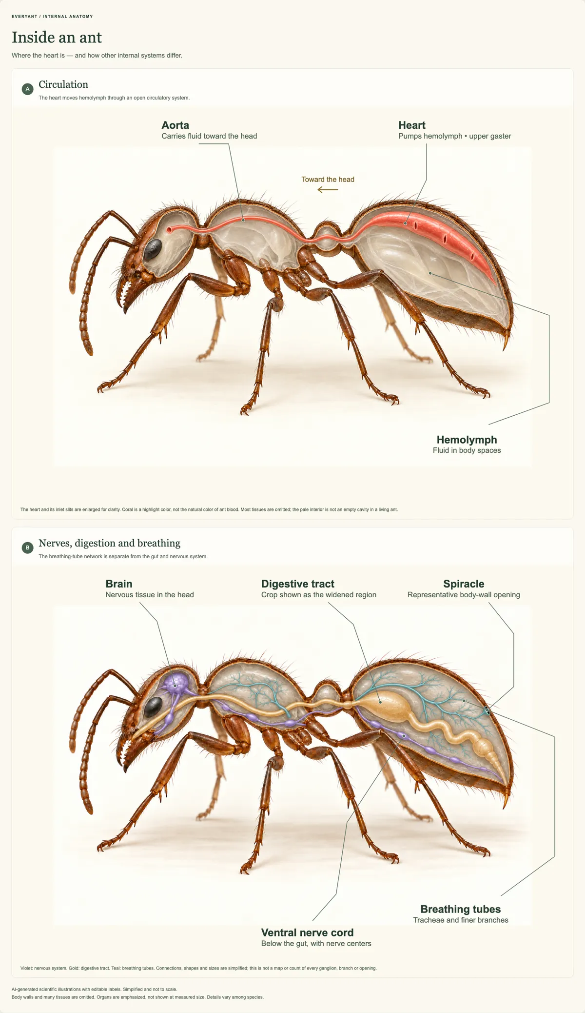
Selected internal systems of an adult worker ant. The heart circulates hemolymph, while a separate network of breathing tubes supplies oxygen. AI-generated illustrations with editorial labels; simplified and not to scale. Body walls and many tissues are omitted, and selected organs are enlarged or emphasized for clarity. Colors are illustrative; details vary among species.
Do ants have blood—and how do they get oxygen?
People sometimes call an insect’s circulating fluid “blood,” but hemolymph is the more precise term. It transports dissolved substances and contains cells involved in protection and repair. Its role extends beyond simply moving food around the body. NC State
Oxygen takes a different route. Air enters through small openings called spiracles and passes into branching tubes called tracheae. Their finer branches bring oxygen close to the tissues that need it.
Ants therefore do not need a miniature pair of lungs attached to a human-style blood circuit. Their breathing system distributes air through the body, while their circulatory system moves hemolymph. Keeping those two pathways separate makes the internal diagram much easier to understand. NC State: respiratory system
Do ants have hearts and brains?
Yes—an individual ant has both. The heart pumps circulatory fluid, while the brain processes information as part of the nervous system.
The brain sits in the head and connects to a ventral nerve cord, running along the lower side of the body. Associated clusters of nerve cells, called ganglia, help process signals and control activity. The brain is consequently part of a distributed nervous system, rather than the only place where information is handled. NC State: nervous system
This anatomy belongs to each ant. It is different from the coordinated behavior of thousands of nestmates. An ant colony does not have one shared physical brain; our colony-biology guide explains how workers’ interactions produce organized colony behavior.
What other organs do ants have?
Ants have organs for processing food, removing wastes and reproduction. Their small size does not mean the inside of the body is an undifferentiated space.
Digestion and food storage
The digestive tract runs from the mouth to the rear of the body. Different regions handle different tasks.
The crop is a food-storage region of the foregut. Farther along, the midgut is an important site of digestion and nutrient absorption. The hindgut processes material before it leaves the body and also helps regulate water and salts. This is the general insect arrangement; the details of feeding structures vary. NC State: digestive system
Removing wastes
Insects also have Malpighian tubules: slender structures that transfer wastes from hemolymph into the digestive tract for elimination.
They work alongside the hindgut’s water- and salt-regulating functions. Removing waste and conserving water are therefore connected processes, rather than separate jobs performed by miniature versions of human kidneys. NC State: excretory system
Reproduction
Reproductive anatomy differs between queens, workers and males, and between species. A queen’s gaster contains her reproductive system, including ovaries that produce eggs. ASU
Workers should not be drawn with a queen’s fully developed reproductive system. Some have ovaries with limited reproductive capacity, some can reproduce under particular conditions, and others lack ovaries altogether. Research comparing worker anatomy across ant species documents this variation. Worker reproductive-anatomy study
For the colony-level explanation, see our guides to queen ants and nuptial flights.
Can scientists measure an ant’s heartbeat?
Yes. In a study published in 2021, researchers developed a method using a modified light microscope and infrared light to record cardiac contractions without destroying the ant. They obtained baseline heart-rate data from three ant species collected in Georgia, USA. Davis and colleagues
The study demonstrates that ant heartbeats can be observed and measured. It does not establish one normal rate for every ant. A measurement belongs to the species, individual and conditions being studied.
Watching an ant walk across a path cannot reveal that measurement. Researchers need an appropriate view of the internal pumping structure and a controlled recording method; ordinary movement of the ant’s body is not, by itself, evidence of its heartbeat.
Further reading
Sources and review notes
EveryAnt prioritizes peer-reviewed, university, museum and government sources. Internal source checks do not replace independent specialist review.
- Ant AnatomyArizona State University · Accessed 2026-10-16
- The Insect Circulatory System: Structure, Function, and EvolutionHillyer and Pass, Annual Review of Entomology (2020) · Accessed 2026-10-16
- Circulatory SystemNC State University · Accessed 2026-10-16
- Respiratory SystemNC State University · Accessed 2026-10-16
- Nervous SystemNC State University · Accessed 2026-10-16
- Digestive and Excretory SystemsNC State University · Accessed 2026-10-16
- What the Heart Looks LikeNational Heart, Lung, and Blood Institute · Accessed 2026-10-16
- How to Take an Ant’s PulseDavis, Clancy and Sasaki (2021) · Accessed 2026-10-16
- Degeneration Patterns of the Worker Spermatheca During Morphogenesis in AntsGotoh, Billen, Hashim and Ito (2016) · Accessed 2026-10-16
- Ultramorphology and Histology of the Dorsal Vessel — Gastral Portion — of Camponotus rufipesMarques and Caetano, Sociobiology (2001) · Accessed 2026-10-16
Published abstract consulted; the full paper was unavailable. Supports middorsal position and slit-like ostia in the studied species.
- X-Ray Microtomography for Ant TaxonomyHita Garcia and colleagues, PLOS ONE (2017) · Accessed 2026-10-16
Artwork context: internal brain, nervous and digestive structures are visible, but the authors warn that dried specimens distort soft tissues. The illustration is not traced from these scans.